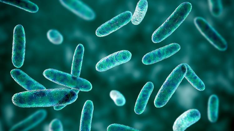
Krakow scientists develop world's first method for identifying bacteria and fungi in microscope images
 Credit: Adobe Stock
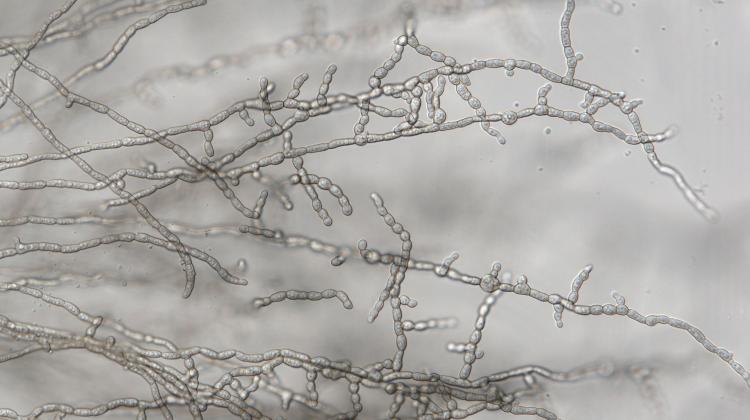
Jagiellonian University scientists have developed the world's first method of fast identification of bacteria and fungi in photographs from light microscopes. The new technology, intended to support medical diagnostics, can also be applied in industry and food safety monitoring.
The new technology utilises deep neural networks and artificial intelligence. The university wants to introduce it to clinical use as quickly as possible.
According to the Centre for Technology Transfer CITTRU of the Jagiellonian University, the use of neural networks and artificial intelligence to analyse images from light microscopes makes it possible to quickly generate a report with a list of identified microorganism species. On the one hand, this solution will significantly speed up diagnostic tests and the implementation of appropriate therapies, and on the other - it will help confirm diagnoses and identification of microorganisms carried out with traditional laboratory methods.
The technology was developed at the Jagiellonian University in collaboration with the Department of Molecular Medical Microbiology, coordinated by Professor Monika Brzychczy-Włoch, and the Institute of Computer Science and Computer Mathematics, coordinated by Dr. Bartosz Zieliński, a professor at the Jagiellonian University.
'The user only has to upload microscopic images of microorganisms to the computer system. In response, the system generates a report with a list of specific species of bacteria present in the analysed material. The entire operation takes no longer than one minute,’ says Dr. Zieliński.
Although the entire procedure only takes a short while, it is very complex. It is based on deep neural networks and nearly 10 years of interdisciplinary research conducted at the Jagiellonian University.
'It all started with an idea put forward a few years ago, to try to combine the forces of computer scientists and microbiologists,' says Professor Brzychczy-Włoch. 'We asked ourselves whether it would be possible to teach artificial intelligence to recognize specific species of bacteria in images seen under a microscope. Fortunately, computer scientists have taken up this challenge and today we have technology that is ready to be tested in a clinical setting.’
The new solution works thanks to specially optimised deep neural networks that analyse microscopic images representing species of both bacteria and yeast-like fungi to detect and precisely identify microorganisms in biological material.
Dr. Zieliński emphasises that one of the challenges consisted in adapting neural networks to effectively analyse a limited amount of input data. Neural networks typically work on huge amounts of data, while here the scientists only had a relatively small amount of data at their disposal, in the form of clinical images and photographs of microbiological cultures. This required a number of measures taken to adapt neural networks to this specific task.
'Our first research was relatively easy and progressed quickly. Especially when the solution was tasked with identifying microorganisms based on photographs from the cultivation of specific bacteria or fungi,’ says Dr. Zieliński. 'However, problems appear when the catalogue of microorganisms that the system can recognize expands, and microscopic images show high densities of many types of bacteria. In such situations, species identification is difficult, and impossible for the human eye. I consider refining the algorithms at this level and achieving a satisfactory level of prediction, i.e. exceeding 90%, to be our considerable achievement.'
The researcher emphasises that an important advantage of the solution is that it make it possible to identify specific species based on the analysis of the image of the morphology of microorganisms. This condition is met even if the material contains several types of bacteria.
Currently, the system can identify several dozen species of bacteria and yeast. According to the scientists who developed the solution, there are no limitations to its future development and diagnostic capabilities. Ultimately, the system should be able to recognise any species with which it has already become familiar. To that end, the scientists have began building the microbe database from the most commonly found pathogens. At the following stages they will teach the system to recognise rarer species.
The authors of the system point out that it can successfully identify not only bacteria and fungi, but also protozoa, for instance unicellular malaria protozoa. It could offer an interesting alternative to relatively costly diagnostic methods, such as Real-time PCR or MALDI TOF, which, additionally, do not yield immediate results.
In addition to clinical applications, the new solution can also prove useful in other areas where microbes need to be detected and identified by means of microscopy.
'Our system may ultimately function as an independent and autonomous tool for identifying microorganisms in the tested biological materials; it may also support or complement previously used laboratory methods. It all depends on the model adopted in the laboratory. Importantly, the test result is delivered almost immediately after the photo is uploaded to the system. This is a key issue in situations where the patient has already developed an infection and a quick diagnosis is needed to immediately apply appropriate treatment,’ says Professor Brzychczy-Włoch.
The Jagiellonian University would like to introduce the new technology into clinical use as quickly as possible. However, for this to happen, further research using clinical biological materials are necessary and a number of procedures must be completed. This requires an industry partner and additional financing.
'Of course, we do not rule out financing research work with public or EU funds, but it is important to improve the technological capacity of this method with an experienced industry partner. This will definitely accelerate commercialisation,’ says Dr. Gabriela Konopka-Cupiał, Director of the Centre for Technology Transfer CITTRU of the Jagiellonian University.
The solution has already been awarded patent protection in the European Union. The creators emphasise that at the stage of patentability testing it was shown that this was the first such technology developed in the world.
PAP - Science in Poland, Katarzyna Czechowicz
kap/ agt/
tr. RL
Przed dodaniem komentarza prosimy o zapoznanie z Regulaminem forum serwisu Nauka w Polsce.