Cells soften or harden as cancer approaches which can be used in diagnostics, say scientists
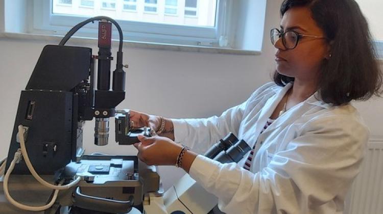 Dr. Kajangi Gnanachandran (Institute of Nuclear Physics PAS) assembles the AFM components used to measure the mechanical properties of cells. Credit: Institute of Nuclear Physics PAS
When healthy cells transform into cancer cells, their mechanical properties change. An international group of scientists, including researchers from Poland, wants to create a procedure for detecting disorders in the mechanical properties of cells in order to diagnose cancer early.
This is an important step towards a novel method of early cancer diagnosis; instead of waiting several weeks for the result, the patient will receive it after a few days, say experts from the Institute of Nuclear Physics of the Polish Academy of Sciences in Krakow.
In 1999, scientists from the institute showed that the cytoskeleton of cancer cells is more 'deformable' than that of healthy cells. This makes it easier for them to squeeze through narrow vessels of the circulatory or lymphatic system and form metastases. Researchers now know that breast, intestinal, bladder and prostate cancer cells become softer already in the early stages of cancer transformation, while cells of other cancers, such as leukaemia, become harder.
Changes in the mechanical properties of cells may also be caused by other factors, such as inflammation. However, it predisposes the patient to further, more precise tests.
'If we had a repeatable measurement procedure, we could use appropriate laboratory equipment to quickly detect disorders in the mechanical properties of cells, which strongly indicate the possibility of cancer developing in the patient's body. The term +quickly+ has two meanings here. On the one hand, we can try to diagnose the risk of cancer at such an early stage of its development, at which other tests usually do not show significant cellular changes. On the other hand, the point is simply that the measurement procedure itself is not very troublesome, it does not require large amounts of biological material and does not take much time,’ says Professor Małgorzata Lekka from the Institute of Nuclear Physics PAS.
Changes in the biomechanical properties of cells can be measured using atomic force microscopes (AFM). AFMs are generally used to image the microworld, even at scales that enable the detection of single atoms. Their probes make it possible to apply a precisely determined force to the tested substrate. If the substrate is a cell, its mechanical response allows the scientists to determine the elasticity coefficient (Young's modulus) and, on this basis, draw conclusions about the elasticity of the structures not only at the cell membrane, but even near the cell nucleus.
According to the Institute of Nuclear Physics PAS, atomic force microscopes are not the most expensive laboratory equipment, but they are not cheap either. Fortunately, there are simplified versions of them: devices called indenters, without imaging functions, but fully sufficient to study the mechanical properties of cells.
'The main factor that has so far limited the development of our method of diagnosing the risk of cancer has not been the cost of the equipment, but the lack of an appropriate measurement procedure. To put it simply, the results obtained in different laboratories, on equipment from different manufacturers, on differently prepared samples, were not repeatable enough to make responsible decisions on further medical action,’ says Professor Lekka. Therefore, standardization of measurements is necessary.
The scientific journal Nanoscale published an article prepared as a result of several years of collaboration of European scientists from universities in Amsterdam, Barcelona, Bremen, Lille, Marseille, Milan, Münster and the Institute of Nuclear Physics PAS in Kraków. An international group of researchers shows that by following a carefully developed procedure, the same Young's modulus value will always be obtained for the same cells, regardless of the place of measurement or the manufacturer of the equipment. The procedure protocol includes, among other things: preparation of samples, calibration of measuring equipment and method of analysing the results. To increase the reliability of the measurements, it was critical to take into account the influence of the substrate, on which the cancer cells were deposited.
'Changes in the mechanical properties of cells occur earlier than optical changes in cancer, so the proposed method will enable the disease to be detected further in advance than before. The value of this headway will probably vary between different types of cancer (...) The new diagnostic method is more sensitive than the optical techniques currently used in cancer diagnosis. The use of standardized measurement procedures, together with automatic data recording and analysis, will allow the test to be carried out in a shorter time. Instead of waiting several weeks for the result, the patient will be able to receive it after just a few days,’ the authors of the publication believe.
The scientists intend to focus on further reducing the number of false positive diagnoses and on testing the procedure in selected disease entities. Before the technique of mechanical detection of cancer lesions reaches hospitals, a stage of clinical trials carried out in cooperation with interested medical units will be necessary.
Research work on the mechanics of cancer cells was carried out as part of the Phys2BioMed project, financed by a Maria Skłodowska-Curie grant from the European Union's Horizon 2020 programme. (PAP)
PAP - Science in Poland
kol/ bar/ KAP/
tr. RL
Przed dodaniem komentarza prosimy o zapoznanie z Regulaminem forum serwisu Nauka w Polsce.