Image plays a huge role in our culture. As a result, the media constantly feeds us graphics of the SARS-CoV-2 coronavirus, which show round balls with coloured spikes. But are these visualizations consistent with what we really know about the appearance of the coronavirus?
According to virologist Professor Krystyna Bieńkowska-Szewczyk from the University of Gdańsk, the answer is yes and no.
The scientist says that although some visualisations are the work of 3D graphic artists, others are indeed based on what we actually know about the structure of the coronavirus.
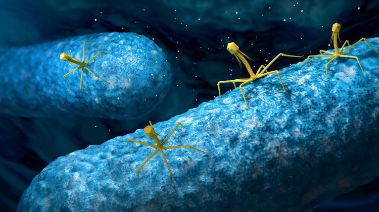
The shape of SARS-CoV-2 - a round structure with spikes - is generally consistent with the results of microscopic observations.
She told PAP: “Thanks to the images from electron microscopes, we know that the coronavirus structure includes a characteristic corona, resembling a luminous shell visible during a solar eclipse.
“This crown is formed by protruding particles on the surface of the virus, called spike proteins. This protein interacts with the host cell and serves as a key to open it.”
But she adds that not everything should be taken literally in the images of the coronaviruses we encounter. The colours of pathogens should be treated as conventional in these images.
She said: “The images from the electron microscope are black and white. But black does not mean that a given structure absorbs light, and white reflects it. In an electron microscope it is not a beam of light that is sent towards the object, but electrons. They fall on a specific object and either pass through it or are reflected from it.
“For example, in classical electron microscopy, objects are covered with a salt of heavy metals that add contrast to the image. So it is difficult to talk about the colours of the sample.”
She added: “Coronaviruses are too small to see them under an optical microscope. Only larger objects, such as bacteria or cells, can be observed.”
Any images that depict coloured coronaviruses (usually there are red or purple parts) must be treated as conventional. In electron microscope photos, colour is added as an element of image processing to make the image more pleasing to the eye, or to make it easier for recipients to distinguish different coronavirus structures or to pick it from the background.
The virologist explained that nano-object imaging techniques are constantly evolving. She said: “X-ray crystallography, electron microscopy, cryomicroscopy are techniques that bring us closer to visualizing real viral structures.”
But these aren't the only ways to experimentally confirm that the coronavirus exists.
Professor Bieńkowska-Szewczyk said: “Evidence for the existence of viruses can also be seen with the naked eye, in virus cultures.
“The virus can only multiply inside a living host cell. Just like a program that only becomes operational in a computer, a virus can only multiply after entering a living cell of a specific type.”
“Where the virus has destroyed cells, it creates a hole in the cell layer. You can even see it with the naked eye.
“While we cannot see the virus itself this way, we can see its effects.
“Another method of watching where the viruses are involves using antibodies. They recognize a given virus, specific viral proteins. A colour factor can be attached to antibodies. Antibodies bind to the virus and the colour reveals its location. In fact, this means that the entire cells that reproduce the virus will be stained and we can count them.”
Bieńkowska-Szewczyk’s team uses these different imaging methods to work with many viruses, such as hepatitis C, Zika or the flu, but not yet with the coronavirus.
She said: “To work with the human coronavirus, we need to modify the laboratory, because it is an airborne virus. To be able to work with such a pathogen, the laboratory and all employees must be properly secured to prevent the virus from being taken out of the laboratory.”
She added: “Although coronaviruses are tiny, there are many ways to see them. The resolution we obtain is getting better. With the help of modern technologies, we are able to reproduce the structure of the viral particle more and more perfectly.”
(PAP)
Author: Ludwika Tomala
lt/ agt/ kap/
tr. RL
Przed dodaniem komentarza prosimy o zapoznanie z Regulaminem forum serwisu Nauka w Polsce.