In a healthy eye, the photoreceptor cells slightly change their length when there are flashes of light. A Polish team is developing a method to record these nano-changes. As a result, it will be possible to precisely image the work of the eye and check whether everything is working properly.
Thanks to a series of breakthroughs in science and medicine in recent years, huge opportunities are opening up for humanity when it comes to the treatment of eye diseases. According to eminent eye researcher Professor Krzysztof Palczewski from the University of California, Irvine, within a decade we will probably be able to heal 99 percent of diseases that lead to blindness.
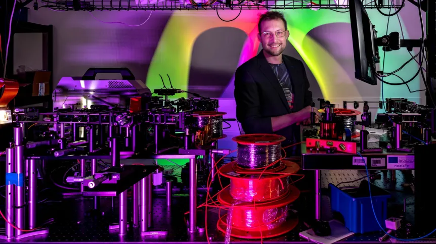
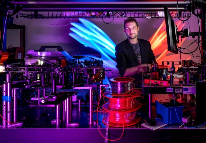
'Introducing new therapies in a short time requires not only biological and medical work, but also new technologies, including those related to eye imaging', says Professor Maciej Wojtkowski, head of the International Centre for Translational Eye Research ICTER, a research agency at the Institute of Physical Chemistry of the Polish Academy of Sciences (Professor Krzysztof Palczewski is also involved in the work of this unit).
As new opportunities to improve vision appear, it will now be more important than ever to diagnose abnormalities in the functioning of the eye early, as well as systematically monitor how the eye subjected to innovative therapies works.
'However, there are still deficits in eye imaging. As a result, new therapies that are being developed cannot be introduced as quickly as doctors would like. There is no way to predict the effects of therapy,’ says Professor Maciej Wojtkowski.
The main goal is to understand the activity of photoactive cells in the eye. We have two types of such photosensitive cells. Rods are more sensitive and process information about the intensity of light (whether it is bright or dim), and cones provide information about colours (there are three types of cones: responsible for seeing blue, green and red light). It is in the photoreceptors that the processes begin to take place, thanks to which we convert the information carried by photons about the outside world into electrical signals that the brain understands.
The work of photoreceptors can be currently studied primarily by electroretinography, which measures electrical potentials in the eye, caused by applying electrodes. And then the points from which the signal comes are roughly analysed. 'These examinations are difficult, poorly reproducible, they require good patient cooperation and are performed in only one percent of cases of daily ophthalmological practice,’ says Professor Wojtkowski.
However, a new idea has emerged on how to see the work of photoreceptors. 'An interesting effect was recently identified: when we shine visible light on the retina of the eye, the photoreceptors begin to change their length,’ Wojtkowski continues. There are two mechanisms leading to the resizing of these cells.
'If we completely get used to the dark and then suddenly turn on the light, the elongation of the receptors is very large. You can say that the retina swells for a while.
‘However, this is related to the process of accommodation - the eye begins to adapt from a mode sensitive to a small amount of light to a mode with a lot of light. That is why at night, when you get out of bed and suddenly turn on the light, it's easy to have an accident - the photoreceptors quickly become saturated with light and you cannot see anything,’ Wojtkowski says.
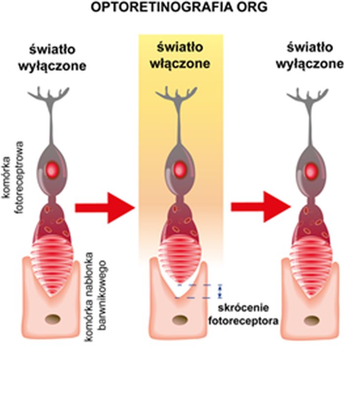
There is another effect. 'When you shine only a very short flash into the eye, the photoreceptors begin to shrink slightly. When it gets dark again, they return to their base length. This process is short and unlikely to be related to the accommodation of the eye, but to the visual cycle. So far, it has not yet been explained why this is happening,’ says Wojtkowski.
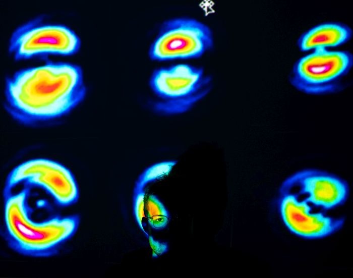
The scientists want to use the latter effect in their idea, which they call optoretinography (ORG). Wojtkowski continues: ‘As part of the study, we shine with short flashes that correspond to the eye's reaction time to light, i.e. with a frequency of less than 50 hertz. And we are watching these very small - nanometer-sized - changes in the length of the photoreceptors. In an instant, we see whether the photoreceptors in the whole eye are responding correctly or not; we will get information about whether the retina is working properly.’
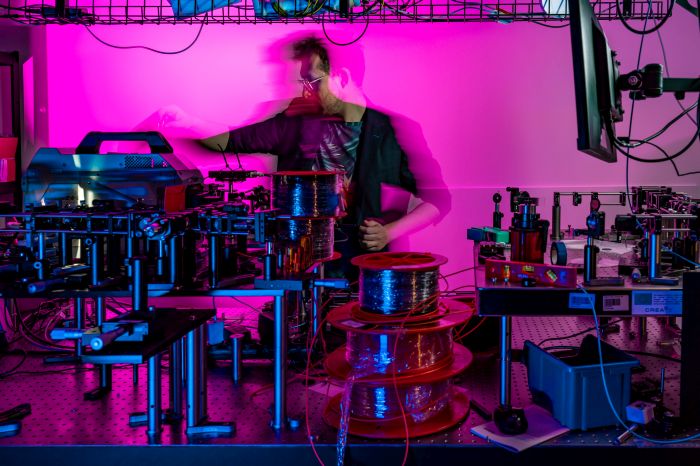
However, in order to collect information about changes in the length of photosensitive cells, scientists had to develop a completely new imaging method based on optical tomography. It is called STOC-T (Spatio-Temporal Optical Coherence Tomography).
'We are looking at whole fundus images taken with a very fast camera. We collect multiple images for different wavelengths. In addition, we introduced spatial decoupling to see the structure of the retina more precisely - to get rid of the side effects related to light scattering, says Professorj Wojtkowski, winner of the Foundation for Polish Science Prize.
How to test it? 'We can distinguish the layer where the photoreceptors start and the layer where they end. We know that where they start - that layer will be the same all the time, and where they end - it will change (shift) under the influence of light,’ Wojtkowski says.
Only the relative difference is analyzed - i.e. the differences between the characteristics of the light emitted by these layers. Wojtkowski continues: 'The trick is that in our method we have access to the light phase. If we can represent light as a sine wave, we can see that the sine representing that wave has shifted even a fraction. Thanks to this, we are able to see even nanometer-sized differences in images. A nanometer is a millionth of a millimeter.’
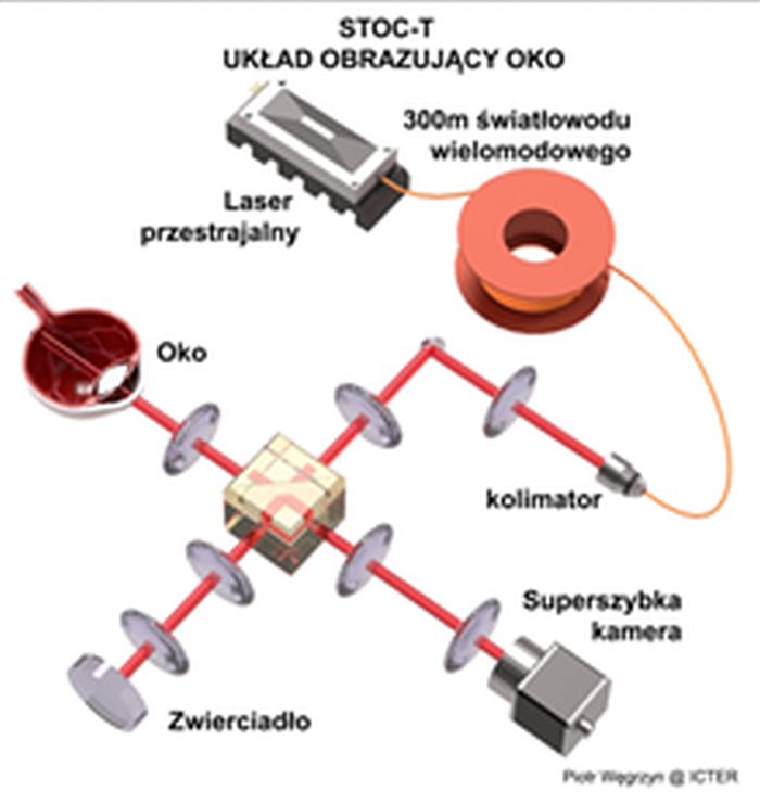
Differences between light phases will be readable even if the patient's eye moves during the examination. The computer can stabilize the image to capture interesting differences. The examination will look like a standard ophthalmological examination - a patient puts the chin on the stand and looks at the device, and the examination takes less than a second. During this time, about 100 measurements are collected, which are then analysed by a computer algorithm.
Professor Maciej Wojtkowski's team expects the prototype of the device to be ready by the end of this year. 'However, we need a few years to develop research, create a database, improve the instrument, refine the software,’ he says.
'Our team is unique in the world because we know how to combine physical skills, optical, technical, IT and biochemical knowledge. This requires a good coordination of groups,’ Professor Wojtkowski concludes. (PAP)
PAP - Science in Poland, Ludwika Tomala
lt/ bar/ kap/
tr. RL