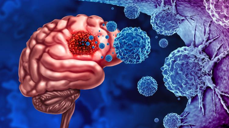
Polish experts develop new, more effective glioma treatment method
 Credit: Adobe Stock
Experts from Bydgoszcz have developed a new method for more precise diagnosis of glioma, allowing for more effective surgery on this extremely dangerous brain tumour.
Glioblastoma is one of the most difficult tumours to treat and operate on. A serious problem is the underestimation of the size of these tumours due to the limited possibilities of brain MRI. The new method involves the use of modified positron computed tomography (PET) imaging.
As reported in Nature Communications, its authors are experts from the Oncology Centre and the 10th Military Clinical Hospital in Bydgoszcz, the Collegium Medicum of the Nicolaus Copernicus University and the Bydgoszcz University of Science and Technology. They claim that it may reach clinical practice within a few years.
'We have shown that in each case we are able to determine glioma infiltration beyond the area visible in MRI. This indicates that even the most radical tumour resection using MRI actually left a significant portion of the tumour behind, resulting in rapid disease recurrence. The discovery is a consequence of our main achievement, which is the verification of another brain imaging technique: PET examination using fluoroethyltyrosine,’ says Professor Maciej Harat from the Bydgoszcz University of Science and Technology, one of the authors of the new research concept and the first author of the paper presenting the discovery.
The expert explains that imaging gliomas using positron emission tomography (PET) begins with administering harmless amounts of the rapidly decaying fluorine radioisotope 18F, introduced into tyrosine molecules - an amino acid deposited in much higher concentrations in cancer cells than in the healthy brain. When the tyrosine molecule is inside the tumour and the radioisotope atom decays, one of the emitted particles will be a positron, the antimatter counterpart of the electron.
Since there are many electrons in the patient's body, the positron quickly hits one of them and annihilates it, turning into energy. Two photons with characteristic energy then escape from the annihilation point in opposite directions. A PET tomograph registers such photon pairs and, based on differences in detection times, determines the place where annihilation occurred, indicating the location of tumour cells.
Professor Harat's research conducted together with Professor Bogdan Małkowski from the Collegium Medicum of the Nicolaus Copernicus University shows that a promising variant of glioma imaging may be supplementing MRI with PET tomography using tyrosine, performed at two time points. So far, attempts to use tyrosine PET as a marker to detect the boundaries of malignant gliomas were performed 20 to 40 minutes after the administration of the radiotracer. Professor Małkowski's assumption that tyrosine molecules can be captured by the tumour earlier turned out to be crucial.
The researchers found that when the test is performed almost immediately after the administration of the tracer, the tumour image is different because tyrosine uptake is particularly intense in the most aggressive tumour cells. Moreover, the image of glioma obtained in this way differs from that obtained using MRI or current PET imaging methods. It was further noted that in some cases, disease progression was visible with the new method up to eight months earlier than with MRI.
'In neuro-oncology, PET tomography is being used more widely at the stage of diagnosis or disease monitoring, but MRI is still used to determine the goal of surgery or radiotherapy. To prove the greater usefulness of PET using tyrosine, we had to not only clearly verify the nature of the cells in places where the images from both methods were so different, but also show that in the early period after tyrosine administration we would detect the most aggressive areas of malignant tumours,’ says Professor Harat.
The usefulness of the new method of imaging gliomas has been confirmed by brain biopsies performed on patients. Based on PET imaging performed at the Oncology Centre in Bydgoszcz, brain tumours were biopsied at the 10th Military Clinical Hospital: after obtaining the patients' consent, small samples were taken from the areas changed in PET and/or MRI.
Detailed analysis of biological material confirmed the significantly greater accuracy of the new method for imaging gliomas. Thanks to this, during surgery or radiotherapy it will be possible not only to remove cancer cells more precisely, but also to spare normal brain tissue.
'The collected evidence will enable us to better plan further clinical trials and hope that once the method is introduced into clinical practice, we will finally improve the results of treatment of these human cancers that are among the ones with the worst prognosis,’ says Professor Harat. (PAP)
zbw/ bar/ kap/
tr. RL
Przed dodaniem komentarza prosimy o zapoznanie z Regulaminem forum serwisu Nauka w Polsce.