Innovative imaging system helps destroy liver cancer
 Source: press materials
An innovative imaging system using high temperature that will help interventional radiologists destroy liver tumours with very high precision has been developed by Evertop specialists and a team from the Silesian University of Technology. The first treatment with the use of the new technology was successfully carried out on August 2 in Warsaw.
Liver cancer is one of the most common causes of death in oncology patients. According to Prof. Olgierd Rowiński, head of the Second Department of Radiology at the Medical University of Warsaw, speed and accuracy are of particular importance in their treatment. Thus, the most effective treatments are minimally invasive interventional oncology procedures with computed tomography, ultrasound or magnetic resonance monitoring. Unfortunately, in the case of some patients these methods are not sufficiently precise - mainly due to chest movements that cause frequent changes in the alignment of organs.
Evertop and Polish scientists have developed an innovative image navigation system that creates a spatial model of the abdominal cavity, taking into account the movements of internal organs and respiratory distortions. Now the image from computed tomography will be combined with the live ultrasound image. A special program overlays this model on the position of the surgical instrument and guides the operator to move it as safely as possible, we read in the release sent to PAP by the company`s representatives.
"The personalized patient model allows for a precise, three-dimensional location of changes in the surrounding of its structures. The surgeon`s field of vision also includes the movement of tools during the surgery. This allows to select the right trajectory, which will lead to the neoplastic change visualized in the model" - says Ewa Piętka, head of the Project Research Team at the Department of Computer Science and Medical Equipment, Faculty of Biomedical Engineering, Silesian University of Technology, quoted in the press release.
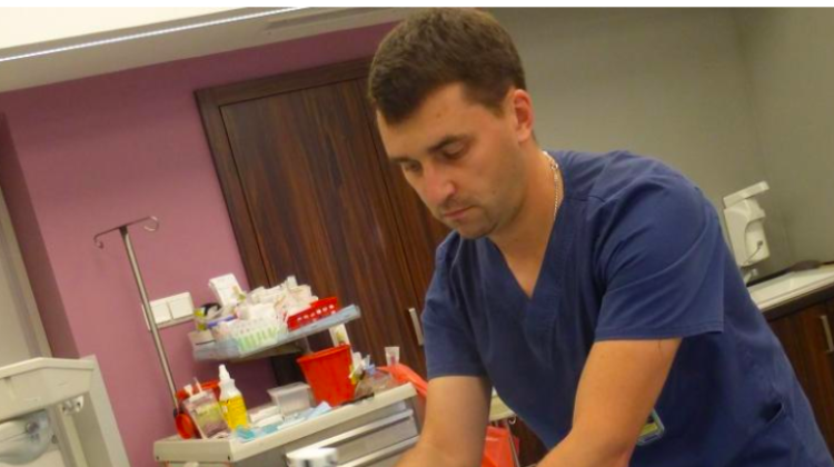
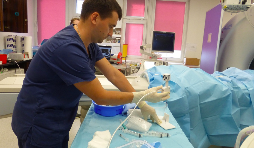
The first treatment with the use of the new technology was carried out on August 2 at the Department of Radiology of the Clinical Hospital in Warsaw (Medical University of Warsaw) in a patient with liver cancer. The next group of patients will benefit from the treatment at the end of the month.
"Liver tumour is our most frequent minimally invasive treatment target in interventional oncology, but sometimes its precise puncture is very difficult. The liver adheres to the diaphragm, so it moves and deforms during breathing" - explains Dr. Krzysztof Milczarek from the Second Department of Radiology, Medical University of Warsaw. "3D imaging, combining real time images, increases the precision of our treatments and reduces the number of complications" - he adds.
The system can be used in all abdominal cavity procedures. In the future, it can also be adapted to treat changes in the chest and pelvis area. Similar solutions are already used in skull and spine surgery, but the mechanisms are much simpler.
"Creating visualization of organs was not difficult, the challenge was the proper reproduction of organ movements and strains during breathing in the algorithms" - explains the project leader Dr. Dominik Spinczyk, from the Silesian University of Technology. "This is the greatest value of our project and we hope that it will revolutionize these treatments in Poland" - he adds.
The project was supported by the National Centre for Research and Development. Evertop received PLN 3 million for the project development as part of the Innomed sector program.
PAP - Science in Poland
ekr/ zan/ kap/
tr. RL
Przed dodaniem komentarza prosimy o zapoznanie z Regulaminem forum serwisu Nauka w Polsce.