Nanoparticles to Light up Early Stage Tumor
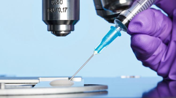 Photo: Fotolia
Nanoparticles that detect tumours and early-stage cancer through a technique called magnetic resonance imaging can replace the current use of kidney-toxic contrast agents.
Animal tests carried out at the Warsaw University of Life Sciences showed that fluorescent nanostructures are effective not only in diagnostics, but also in biopsies.
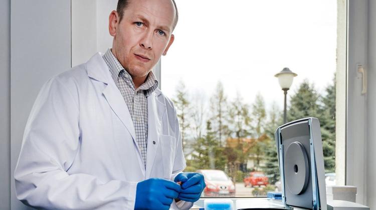
A team of scientists led by Dr. Michał Godlewski from the Department of Physiological Sciences of the Institute of Veterinary Medicine in cooperation with the Veterinary Research Centre have been working on compounds that are so neutral to the body that they can be used in screening.
During magnetic resonance imaging doctors currently use gadolinium as a contrast agent. But although this produces accurate measurements it has a neurotoxic effect on the kidneys. It also accumulates in the nervous system.
Dr. Godlewski said: ”The accumulation of gadolinium in the brain and its negative effect on the functioning of the kidneys (nephrotoxicity) have been confirmed. Information on the potential neurotoxicity of gadolinium appears in scientific publications, hence it is only a matter of time before the use of gadolinium preparations is prohibited or strictly limited. It is still used because for now there is simply no compound that could replace it in resonance.”
In earlier research he created fluorescent nanostructures and found that nanoparticles given to a test animal would pass into each organ and cross the blood-brain barrier, which he said proved “to be a very important feature desirable for creating markers for medicine," he said.
During subsequent studies, scientists found that nanomaterials are both well absorbed and efficiently removed by systems that maintain the biological balance of the body (homeostasis). The research concerned zinc oxide and zirconium which Dr. Godlewski is now working on using them to fight cancer.
He said: ”The tumour environment behaves completely differently than other tissues. We are dealing with a rapid growth of vessels that have a very loose structure. Various substances can penetrate it without any problem. In addition, lymphatic vessels do not form in the tumour, which means that it has no drainage system. This makes tumour labelling easier. Fluorescent and magnetic nanoparticles will reach the tumour and accumulate there. During magnetic resonance imaging it will be easy to see such a cluster; it means that this is where the tumour is.”
He added: ”There are already systems with optical visualization, which shows if we are definitely collecting the target material. Nanoparticles can also be used during surgery to identify the area of healthy tissue and cancer infiltration. This is especially important in the case of brain tumours. The brain tumour is cut out, but not completely: a part of it is left, especially in cases where it invades the vital areas of the brain. Like with normal tissue, we cannot afford to remove a large area. In the brain, this margin is necessarily very small.”
Test are still being carried out to ensure that using nanoparticles are not harmful but Dr. Godlewski said: “So far, all the studies that we conduct show that with the doses used (within the range of acceptable European standards of daily intake of the substance), there were no side effects. This was confirmed in studies on laboratory mice.”
There is still a long way to go before applications of nanoparticles can be used in human diagnostics, especially since bioethics committees in Poland have not approved such studies on humans.
In addition to legal problems, there are also financial problems with enterprises reluctant to invest in clinical trials.
Scientists say they are now trying to find investors.
PAP - Science in Poland, Karolina Duszczyk
kol/ ekr/ kap/
tr. RL
Przed dodaniem komentarza prosimy o zapoznanie z Regulaminem forum serwisu Nauka w Polsce.