Printed 3D heart models help in complex surgeries
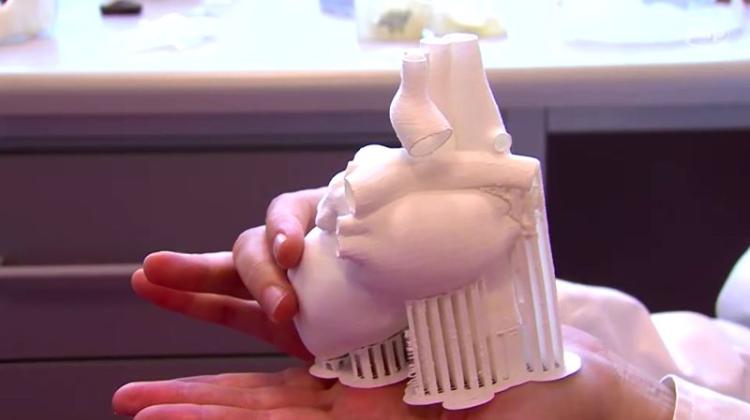
Printed 3D models of the heart or blood vessels are being made at the Laboratory of Individual Medical Implants in Bionanopark in Łódź. Models reflect the heart of a particular patient in detail and help the cardiac surgeons prepare for complicated surgeries.
Using such models, surgeons can practice the procedure, more precisely plan its course and prepare the necessary equipment for the operation" - told PAP Dr. Marcin Elgalal, head of the laboratory.
The Łódź Laboratory of Individual Medical Implants has been making tailor-made implants for several years. These implants allow to reconstruct an orbital bone or skull bones in patients who have lost them, for example due to an accident or cancer.
Based on imaging studies - computer tomography and magnetic resonance imaging - virtual models of various anatomical structures and pathological changes are generated. Computer-generated design may include, for example, bone defects, tumours, vascular pathologies that occur in a particular patient. Based on this virtual model, three-dimensional physical models are printed.
"The surgeon receives, for example, an anatomical model of the orbit and on that basis forms a titanium mesh implant, which is then implanted in the patient. The model can also serve as a training aid and aid during surgery" - Elgalal explained.
The same technology can be used to make soft tissue models, including models of the heart or blood vessels, that serve as tools for planning complicated and difficult operations.
As in the case of hard tissues, the first step is computed tomography. Since the heart model should be very precise, tomography is performed done in such a way as to obtain very accurate data.
"The scan is performed with a very thin layer, with modern, multi-row tomography equipment. Later comes a long, hard work to get all the anatomical details out of this data and create a 3D model of an anatomical structure, in this case the heart, the ventricles, the main vessels connected to the heart" - told the head of the lab.
Models can be printed from hard materials on commonly used 3D printers or from more expensive soft materials on state-of-the-art equipment. The latter models are structurally similar to soft tissues, which in the case of some procedures is very much needed by surgeons.
Such heart models can be used to plan more difficult, more complicated procedures such as congenital heart defects treatment or interventional cardiology procedures. "Surgeons can practice the procedure before surgery. The whole team can gather and look at the model, take it in hand. This makes it a bit easier to imagine the pathologies that occur in the patient, to plan the course of the surgery or the necessary equipment" - Dr. Elgalal emphasised.
The Łódź Laboratory of Individual Medical Implants has prepared only a few such heart models. According to its head, this method will be increasingly used in medicine, because 3D printing offers the opportunity to create accurate anatomical models. "If you look at medical literature, it is evident that such anatomical models are increasingly used for a variety of procedures and operations" - he said.
PAP - Science and Scholarship in Poland
szu/ ksk/ kap/
tr. RL
Przed dodaniem komentarza prosimy o zapoznanie z Regulaminem forum serwisu Nauka w Polsce.