New method for faster testing of tissues and cells

A new method for microscopic imaging and measurement, allowing for faster testing of tissues and cells, is being developed by Polish scientists. With this method, histological analysis will be several times faster, because, for example, staining samples will not be necessary. It will also be easier to examine the effects of new drugs on cells.
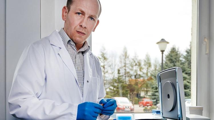
"In histopathology, a tissue taken from the body - for example by means of a biopsy - must be cut, then fixed and stained with appropriate reagents" - Prof. Małgorzata Kujawińska explained in an interview with PAP. Such a long process means that histopathological examination of a single sample may take several days.
The team of Prof. Kujawińska from the Institute of Micromechanics and Photonics, Warsaw University of Technology is working on a new type of microscope - tomographic phase microscope. This device can relieve doctors pathologists, enabling the automation of measuring tissue section without prior staining. The whole measurement process will be faster, simpler and cheaper. "At this point it is difficult to say how much the new method will shorten the test time. It depends on the type of tissue, but I think that the process will be shortened at least a few times, maybe more" - explained Prof. Kujawińska.
When using traditional biological microscope, the best method for accurately measuring the sample is to fix the preparation in paraffin and stain it. As a result of staining, various tissue structures absorb light differently and the colour image viewed under the microscope is interpreted by a physician.
"We say: let\'s do almost the same thing, but skip the colouring process and the introduction of markers. For this we need our tomographic phase microscope. It will allow to analyse without the need for staining preparations" - explained Prof. Kujawińska. "Original tissue structure has a characteristic distribution of the refractive index indifferent locations. We will use tomographic phase microscope to quantitatively analyse the refractive index of the structure" - she described.
Researchers will thus receive an image representing the exact values of the refractive index. Because a colour can be assigned to each numerical value, the data can be visualised, and obtained images virtually stained. "The advantage of tomographic microscope is that if we have a group of cells or thicker tissue, such a structure can be visualized in 3D, because the system will measure the three dimensional distribution of the refractive index in the microstructure" - described the researcher.
In his project, scientists from Warsaw University of Technology propose two versions of the device: for collected samples that remain unchanged and the cultured cells subjected to the influence of, for example, new drugs. "In this scenario, our method will expedite the process of studying the effects of a drugs on a given cell type or tissue. This will accelerate the development process of new drugs" - explained Prof. Kujawińska.
On the other hand, the new device will also useful in so-called tissue plants, allowing to monitor tissue growth and culture for various purposes, for example orthopaedic, cardiac applications. "In the world the number of such +factories+ is growing fast and they lack the tools to control +manufacturing+ processes of biological structures" - emphasised the researcher. The device can also be used to study fundamental processes occurring in biological structures. According to Prof. Kujawińska, interest in this type of tool in research institutions is already very high.
Work is carried out as part of the Foundation for Polish Science grant in the TEAM-TECH programme; researchers have three years to prepare this method. After this period, the devices should be ready for mass production.
PAP - Science and Scholarship in Poland, Ewelina Krajczyńska
ekr/ agt/ mrt/
tr. RL
Przed dodaniem komentarza prosimy o zapoznanie z Regulaminem forum serwisu Nauka w Polsce.